A NEW METHOD OF SIMULATING HUMAN BRAIN IMMUNE CELLS
The research, which was published in Cell on May 11, 2023, emphasises the significance of immune cell and brain interactions and advances knowledge of neurodegenerative and developmental illnesses, including Alzheimer's disease and an autism spectrum disorder. Microglia, specialised brain immune cells, are located at the interface between the human immune system and the brain and are critical in both development and illness. Microglia are unquestionably important, yet modelling and analysing them has remained challenging.
Human microglia are challenging to research when taken out of the human-brain-like environment, in contrast to some human cells that can be examined outside of the body or in nonhuman models. Salk researchers created an organoid model, a three-dimensional collection of cells that resembles the characteristics of human tissues, to get around this obstacle. For the first time, using this model, scientists can examine the growth and operation of human microglia in real human tissue. The researchers also looked at microglia obtained from patients with macrocephalic autism spectrum disorder (a conditionOrganoids, which first appeared about ten years ago, are now a common tool for bridging cell and human investigations.
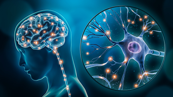
Organoids, as opposed to other laboratory systems, are better able to simulate human growth and organ creation, enabling researchers to explore the effects of medications or illnesses on human cells in a more natural environment. Usually cultivated in culture dishes, brain organoids have morphological and functional limitations due to a lack of blood arteries, a short lifespan, and the inability to support a variety of cell types (like microglia).
"We used a novel transplantation technique to create a human-brain-like environment, to create a brain organoid model that contains mature microglia and enables us to research them," explains co-first author Abed Mansour, a former postdoctoral researcher in Gage's group and currently an academic. Organoids are a popular technique for bridging the gap between cell and human investigations since their introduction about ten years ago. Organoids imitate human growth and organ formation more accurately than other laboratory systems, enabling researchers to explore the effects of medications or illnesses on human cells in a more natural environment.
Typically, culture plates are used to generate brain organoids, however, these organoids have structural and functional limitations because of their lack of blood arteries, short survivability times, and inability to support a variety of cell types (like microglia).
Israeli University's Hebrew University
assistant professor. So that we could ultimately create an organoid of the
human brain with all the qualities required to control human microglia
behaviours, development, and function.
Researchers developed a human brain organoid that is different from earlier versions. They were now able to examine environmental impacts on microglia throughout brain development since it contained microglia and a human brain-like environment. They discovered that the distinctive protein SALL1, which helps to validate microglia identification and enhance mature function, first emerged as early as eleven weeks during development.
Furthermore, they discovered that the activity of microglia was dependent on elements unique to the brain environment, such as the proteins TMEM119 and P2RY12. The development of a human brain model that can accurately mimic the environment of the human brain is highly exciting, according to associate professor Axel Nimmerjahn, another research author. With the help of this model, we can at last look at how human microglia operate in the context of the human brain.
The significance of the connection between the brain environment and microglia emerged as the research team learned more about these cells, particularly in the context of illness situations. In a prior study, the researchers looked at neurons developed by autistic spectrum disorder sufferers and discovered that these neurons expanded more quickly and had more intricate branching than their neurotypical counterparts. The scientists could investigate if such neuronal variations affected the brain environment and impacted microglia growth using the novel organic model. against three macro cephalic people with a neurotypical autism spectrum disorder. The scientists discovered that such neuronal changes were present in people with autism spectrum conditions and that the microglia were affected by those differences in their development environment. The microglia become more reactive to injury or invaders as a result of this neuron-dependent environmental shift, which may help to explain the brain inflammation seen in certain people with autism spectrum conditions.
To confirm their
findings, the team intends to look at more microglia from more persons in the
future, given this was a preliminary study with a limited sample size. To understand how microglia are influencing the start of the illness, they also
plan to broaden their research to examine additional developmental and
neurodegenerative disorders.
They did this by comparing microglia made from skin samples from three different people with We chose to build the brain ourselves," explains Simon Schafer, co-first author and former Gauge lab postbox. Schafer is currently an assistant professor at the Technical University of Munich. We can work from the bottom up and see answers that may be hard to see from the top down by creating our own brain models. We are enthusiastic to keep developing our model and figuring out how the immune system and the brain interact. Other authors include Johannes C. M. Schlachetzki, Addison J. Lana, Christopher K. Glass, Irene Santisteban, Lisa Mitchell, Amanda Mar, Daphne Quang, Sarah Stumpf, and Clara Baek of the Salk Institute; Saeed Ghassemzadeh, Lisa Mitchell, Amanda Mar, and Raghad Zaghal of the Hebrew University of Jerusalem.
Information about the Salk Institute for Biological Studies
The mission of the Salk
Institute is to discover the mysteries of life itself. Our group of top-tier,
outstanding scientists advances knowledge in a variety of fields, including
neurology, cancer research, ageing, immunobiology, plant biology, computational
biology, and more. The Institute is an independent, nonprofit research
organisation and architectural landmark that was founded by Jonas Salk, the man
responsible for creating the first polio vaccine that was both safe and
effective. It is tiny by design, intimate by nature, and brave in the face of
any difficulty. Visit www.salk.edu to find out more. Salk Institute for
Biological Studies information
The Salk Institute's mission
is to discover the mysteries of life itself. In fields including neurology, cancer
research, ageing, immunobiology, plant biology, computational biology, and
others, our team of top-tier, award-winning scientists pushes the limits of
knowledge. The Institute was established by Jonas Salk, who created the first
polio vaccine that was both safe and effective. It is an independent, nonprofit
research organisation and architectural landmark that is tiny by design,
intimate by nature, and courageous in the face of any difficulty. Visit
www.salk.edu to learn more.


Comments
Post a Comment